stylet base conical
Macrostomum coomerensis Fig 4.jpg

the distal two thirds of the stylet are spiralled. The distal tip (of the stylet) is broadend, the opening of which lies subterminally. p. 518
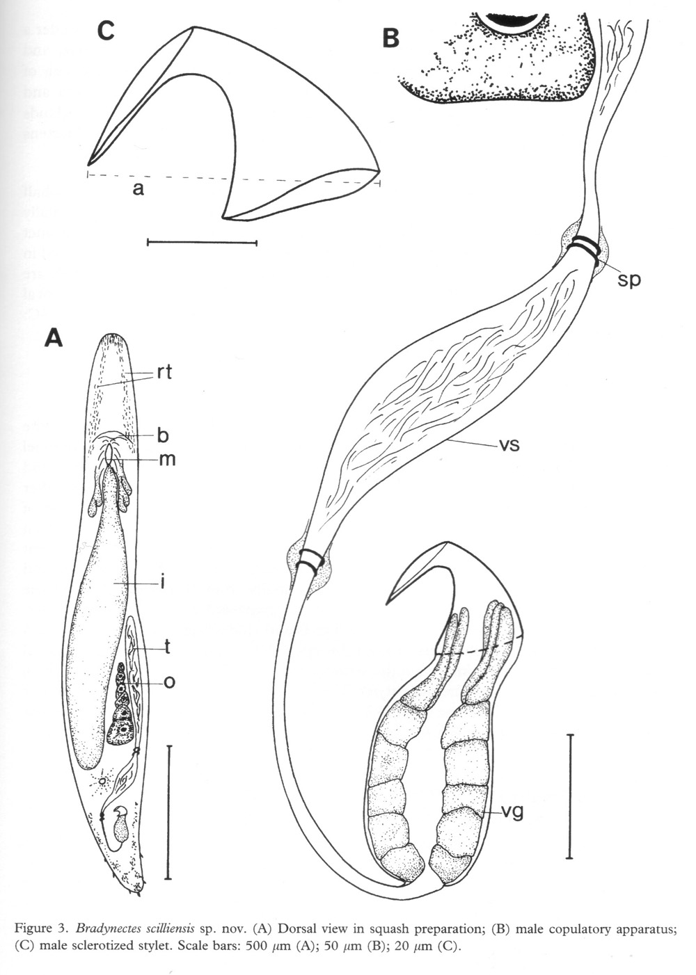
Bradynectes scilliensis Fig 3.jpg
Macrostomum australiense Fig 8.jpg
The stylet is a slightly curved funnel with a widened proximal base and a distal hook-like bend turned at an angle of about 90° from the main axis. p. 1001
Macrostomum virginianum Fig 2-4.jpg
The stilette is curved and obliquely truncated on its convex side. This conical tube is bent at a right angle near its proximal end. p. 27
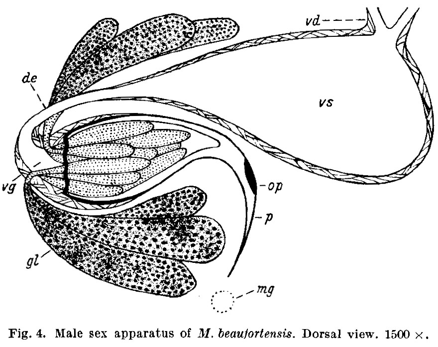
Macrostomum beaufortensis Fig 4.jpg
The stilette is a greatly curved and highly sharpened funnel with a broad undulating basal rim. p. 234
Macrostomum beauchampi Fig 9.jpg
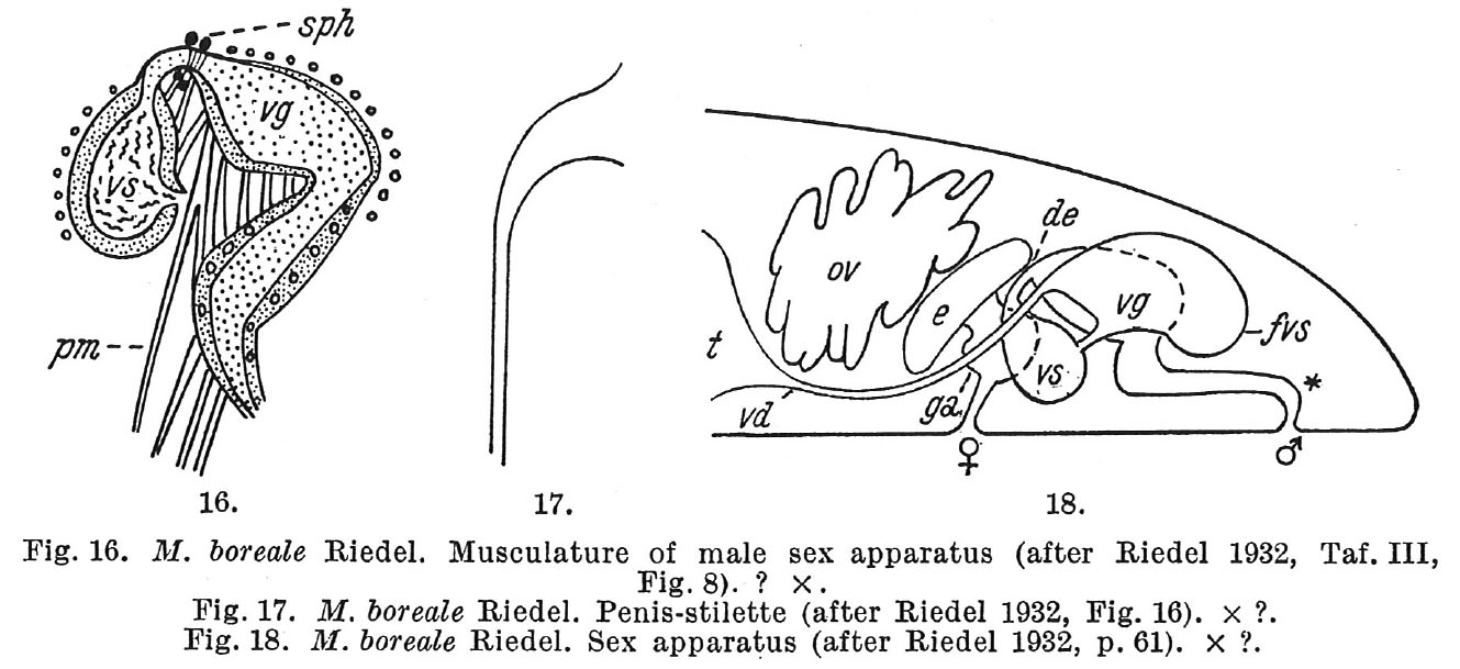
Macrostomum boreale Fig 16-18.jpg
Macrostomum beaufortensis Fig 10-15.jpg
Macrostomum intermedium Fig 24-25.jpg
Macrostomum glochistylum Fig 13-19.jpg
The small reniform eyes are not embedded in the tissues of the ganglia. The penis-stilette is a long, curved funnel which extends from a widened proximal base to end in a slightly bent obliquely truncated terminus. The stilette termination is distinctly provided with a thin lip which projects slightly beyond the opening proper. p. 195 The sperm cell in this form is a spindler-shaped structure which is much shorter than usual for Macrostomum. Chromatin granules are present in the anterior region of the cell, while posteriorly directed setae are absent. p. 196