stylet tip opening terminal
Macrostomum tuba Fig 1-8.jpg

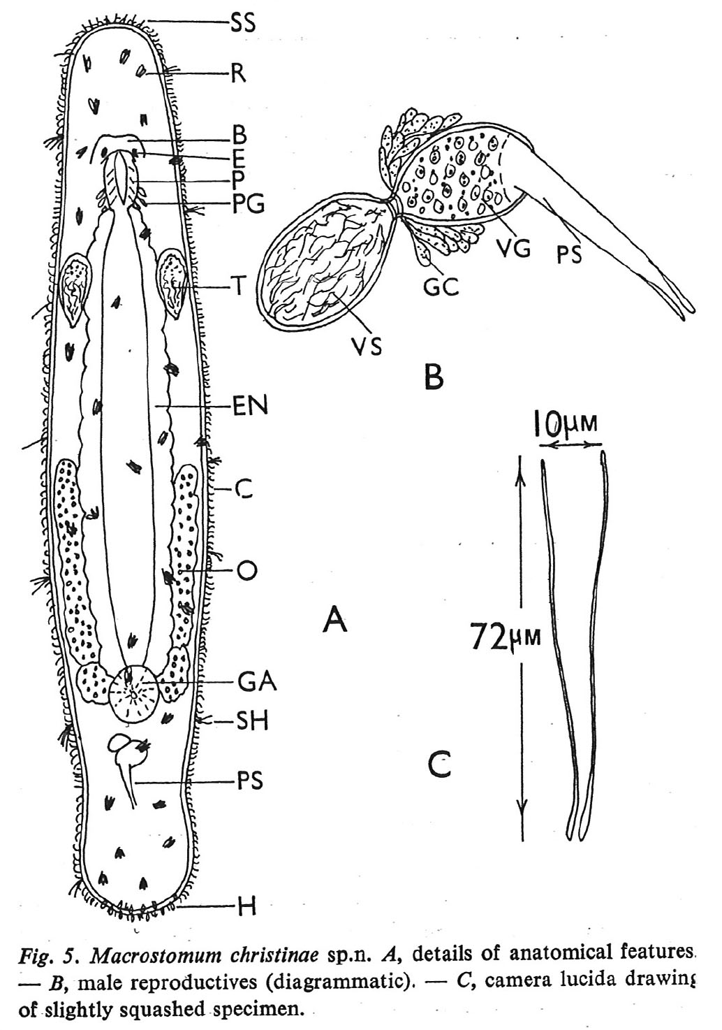
Macrostomum christinae Fig 5.jpg
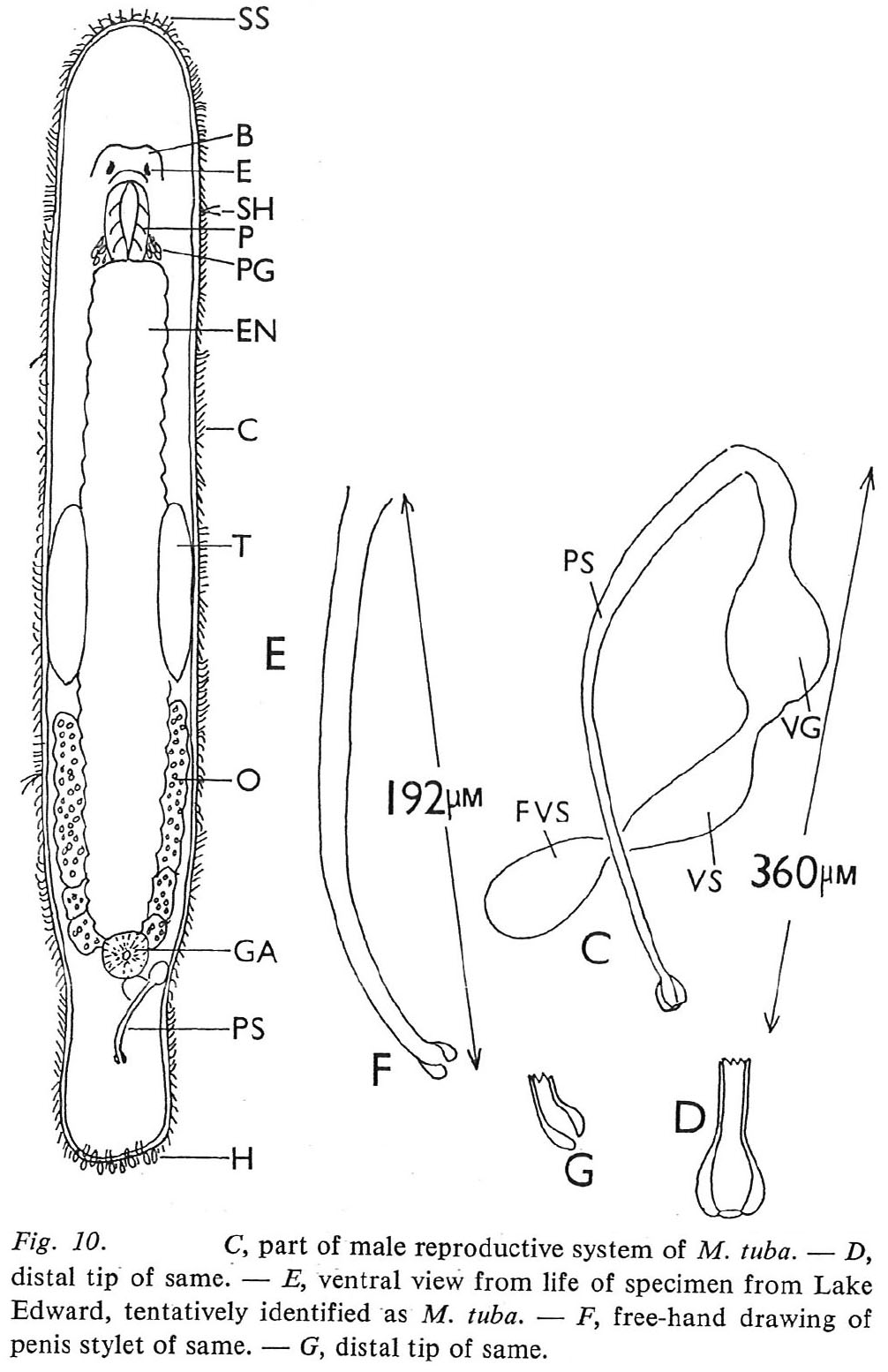
The eyes are small with an irregular shape with dimensions of 8 to 12 micrometer. The penis stylet has its distal end bent at more-or-Iess a right angle to the main tube. This tip constitutes the thickened wall of the tube. The distal opening is situated at the heel of the stylet. p. 51
Macrostomum tuba Fig 10c-d.jpg
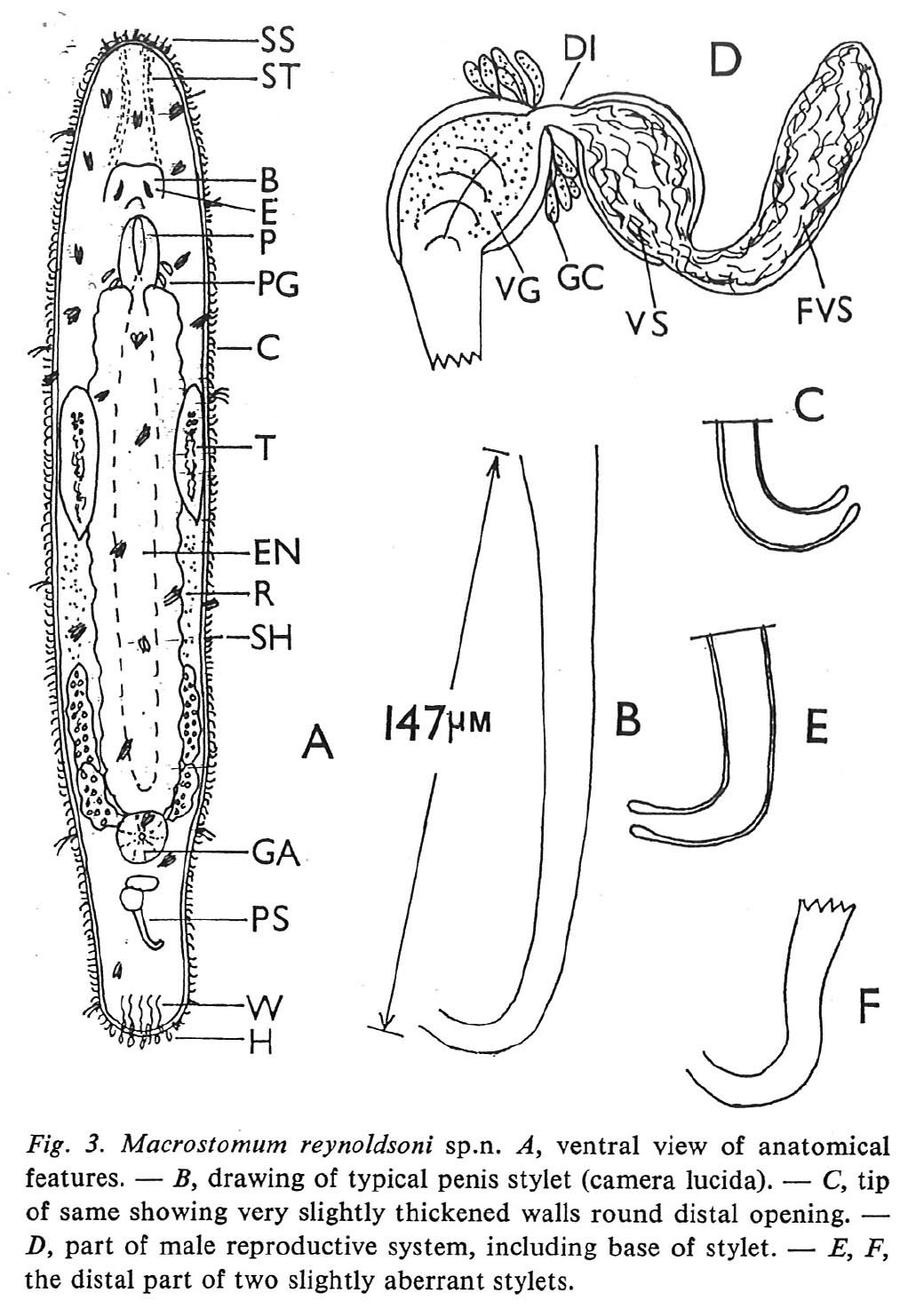
Macrostomum reynoldsoni Fig 3.jpg
The penis stylet is hook-shaped; it is slightly bent in another plane at a point indicated by the arrow. Its walls are not thickened at any particular point. Its distal opening is terminal. p. 53
Macrostomum nairobiense Fig 4.jpg
The penial tube has a slight bend at its proximal end, and a distinct and sharp bend at its distal end. The distal tip of the stylet is blunt with a terminal opening. The walls appeared of uniform thickness along the length of the tube. p. 56
Macrostomum georgeense Fig 8.jpg
The penis stylet is a more-or-less straight tube which diminishes in diameter towards its distal tip; in fact the latter is slightly bent as shown in Figs. 5Band C. The walls of the tube around the distal terminal opening are slightly thickened. p. 54
Macrostomum tuba Fig 3d.jpg
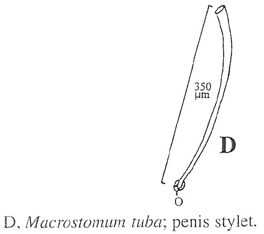
Penis stylet very slightly spiralled; distal end opening superior, i.e. on concave side p. 48
Macrostomum tuba Fig 1.jpg
Macrostomum tuba Fig 40-41.jpg
Von den Spermazellen konnten, allerdings nur am lebenden Objekt, folgende zwei Typen erkannt werden: 1. (Bilder 36 und 37) Der fadenförmige Körper erweitert sich an beiden Enden oval. Das breitere Ende, das bei der Bewegung vorangeht, trägt noch einen fadenförmigen Fortsatz. In der hinteren Erweiterung befinden sich drei bis fünf dunkle Punkte, die vermutlich Chromatinbrocken darstellen. 2. (Bild 39) Beim Typus 2 der Samenzelle trägt der lange fadenförmige Körper nur vorn eine Erweiterung. Sie hat keinen Fortsatz und enthält caudal ein Körperchen, anscheinend den Kern. Die Erweiterung bildet den Kopf des Samenfadens. Der Kopf geht caudal in eine Anschwellung über, die als Mittelstück bezeichnet wird. In diesem liegen zwei kräftige Verdickungen (c), die vermutlich die Zentrosomen sind. p. 176/177